pathology / histopathology

At Cilcare, we specialize in delivering top-tier contract histopathology, immunohistochemistry, and pathology services. Our core expertise lies in the intricate domains of the inner ear and the brain, where precision and depth of knowledge are paramount, but also extends to other organs from multiple species.

histology of the ear

  • Histopathology
    • Paraffin slide preparation (Gross trimming, processing, embedding, sectioning, staining)
    • Evaluation of the external, middle, and inner ear
    • Pathologist reading and scoring
  • Flat surface preparation of the inner ear
    • Immunochemistry & immunofluorescence
    • Cytocochleogram (hair cell counting)
    • Any cochlear structure staining (supporting cells, fibers)
    • Specific markers: ribbon synapses, pre & post synaptic markers
    • Fluorescence monitoring (Gene vector transduction efficiency, fluorescent product)
  • Cross section preparation of the inner ear
    • Paraffin slide preparation (Gross trimming, processing, embedding, sectioning, staining)
    • Immunochemistry & immunofluorescence
    • All cochlear structure staining (hair cells, supporting cells, fibers, SGN)
    • Coloration: H&E (fiber & SGN counting)
    • Fluorescence monitoring (Gene vector transduction efficiency, fluorescent product)

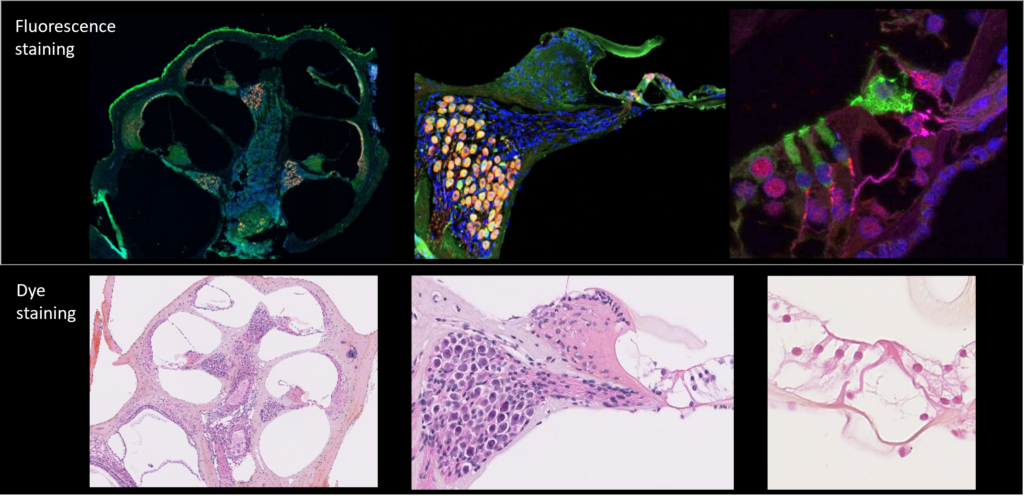
Cochlea cross section

cytocochleogram

Cochlear assessment plays a pivotal role in auditory research. Cilcare’s scientists employ high-level techniques that combine extreme precision, cutting-edge technology, and years of experience to deliver robust and qualitative cochleogram data, whether related to non-GLP or GLP studies.

histology of other organs

  • Liver, kidney, spleen, gonads, heart, lungs, vitreous humor of the eye, Brain, Spinal cord, Bone (femur), Knee
  • Histopathology 
    • Immunochemistry & immunofluorescence
    • Coloration: H&E, cresyl violet, Safranin O Fast Green
    • Scoring of inflammation and cartilage degradation

microscopy & imaging

  • Scanning Electron Microscopy (SEM): Morphological analysis
  • Optic microscopy
    • Fiber & SGN counting
    • Histopathology of middle ear and cochlea
  • Confocal fluorescence microscopy
    • Cochleogram
    • Ribbon synapses
  • Transmission Electron Microscopy (TEM): Observation of cellular morphology and structure
SEM of rat cochlea highlighting damages on the first row of outer hair cells after noise exposure